Medical Ultrasounds Deployed Underwater to Non-Invasively Monitor Endangered Coral Reproduction and Health

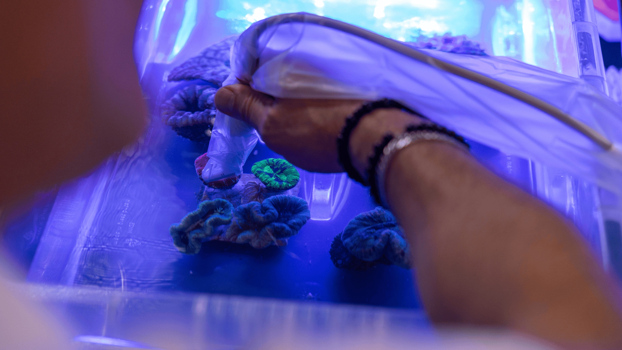
In a novel convergence of human medicine and marine conservation, scientists have turned to an everyday clinical tool—the ultrasound—to diagnose and monitor the reproductive health of endangered coral reefs. Traditionally, assessing whether a coral colony was carrying eggs required destructive sampling, which frequently involved sacrificing or severely damaging the specimen. However, a recent technological breakthrough pioneered by imaging giant Canon in collaboration with United Kingdom-based conservation group Coral Spawning International (CSI) has successfully demonstrated that medical ultrasound platforms can scan living, submerged coral without causing harm.
The breakthrough relies on high-frequency imaging technology originally designed for hospital obstetrics. By adapting these medical devices for marine environments, researchers hope to gain unprecedented insight into coral spawning cycles. This non-invasive diagnostic capability arrives at a critical juncture for marine ecosystems worldwide, offering conservationists a precise new instrument to time and target reef restoration initiatives before time runs out for the world’s most vulnerable marine habitats.
The Mechanics of Underwater Diagnostic Imaging
During a recent series of controlled tests, researchers submerged four distinct living species of coral to evaluate the efficacy of the diagnostic procedure. The multidisciplinary team utilized Canon’s Aplio i700, an advanced medical ultrasound system equipped with specialized high-frequency and ultra-high-frequency probes. While the hardware is typically deployed in clinical settings to monitor human health, its acoustic properties proved remarkably effective when applied underwater to marine invertebrates.
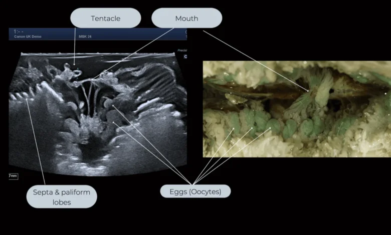
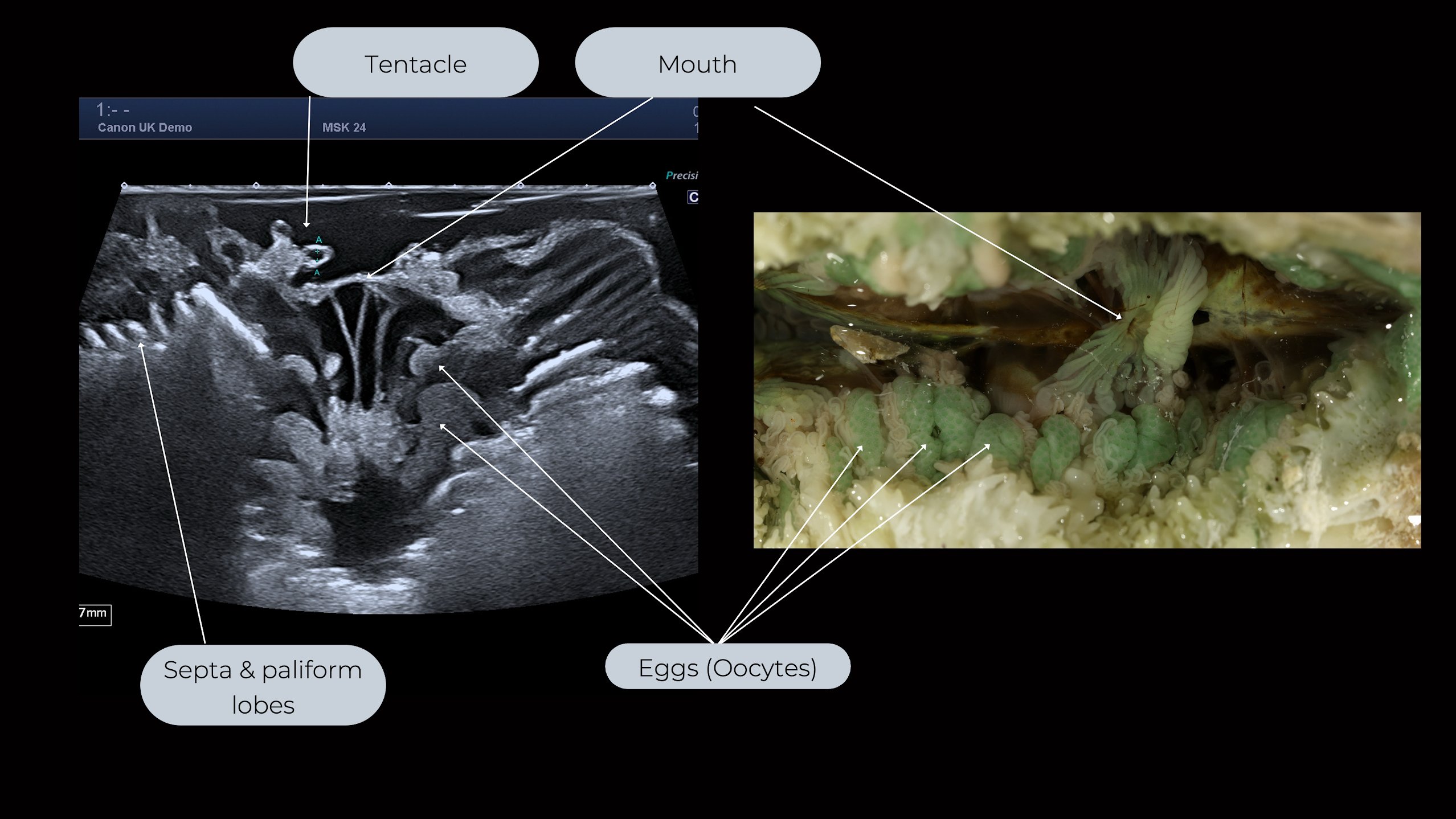
The primary engineering challenge of scanning coral lies in its physical composition. Stony corals possess hard, dense calcium carbonate skeletons that traditionally obscure internal anatomy from external observation. However, the high-resolution beams of the Aplio i700 successfully penetrated these skeletal barriers. The probes mapped the internal architectures of six large polyp stony coral species, clearly revealing tiny circular shapes identified as developing eggs.
To validate the accuracy of the ultrasound data, researchers performed a controlled physical verification on a specimen of Trachyphyllia geoffroyi, a prominent species of large polyp stony coral. By carefully fragmenting and examining the interior of the coral under high-powered laboratory microscopes, scientists cross-referenced the physical anatomy with the prior ultrasound scans. The comparison definitively confirmed that the internal structures highlighted by the ultrasound signals were indeed developing oocytes, or eggs. This verification established the ultrasound platform as a reliable diagnostic tool for non-invasive marine biology.
Chronology of the Innovation and Collaborative Effort
The integration of clinical ultrasound technology into marine science represents months of iterative engineering and field testing between corporate developers and ecological field researchers.
The initiative began as an exploratory venture to determine whether high-frequency sound waves could travel effectively through seawater and penetrate dense skeletal matrices without distortion. Initial laboratory tests in controlled aquariums focused on calibrating probe frequencies to compensate for the acoustic impedance differences between human tissue and marine calcium carbonate.
By late autumn, the partnership between Canon and Coral Spawning International moved from conceptual design to practical application. Field teams conducted trials using portable power units and waterproof housings to operate the medical equipment dockside and in shallow marine enclosures. Following the successful identification of egg structures in Trachyphyllia geoffroyi and related species, the project leaders formally announced the technological milestone to the scientific community, establishing a blueprint for future field deployments.
Official Responses and Corporate Perspectives
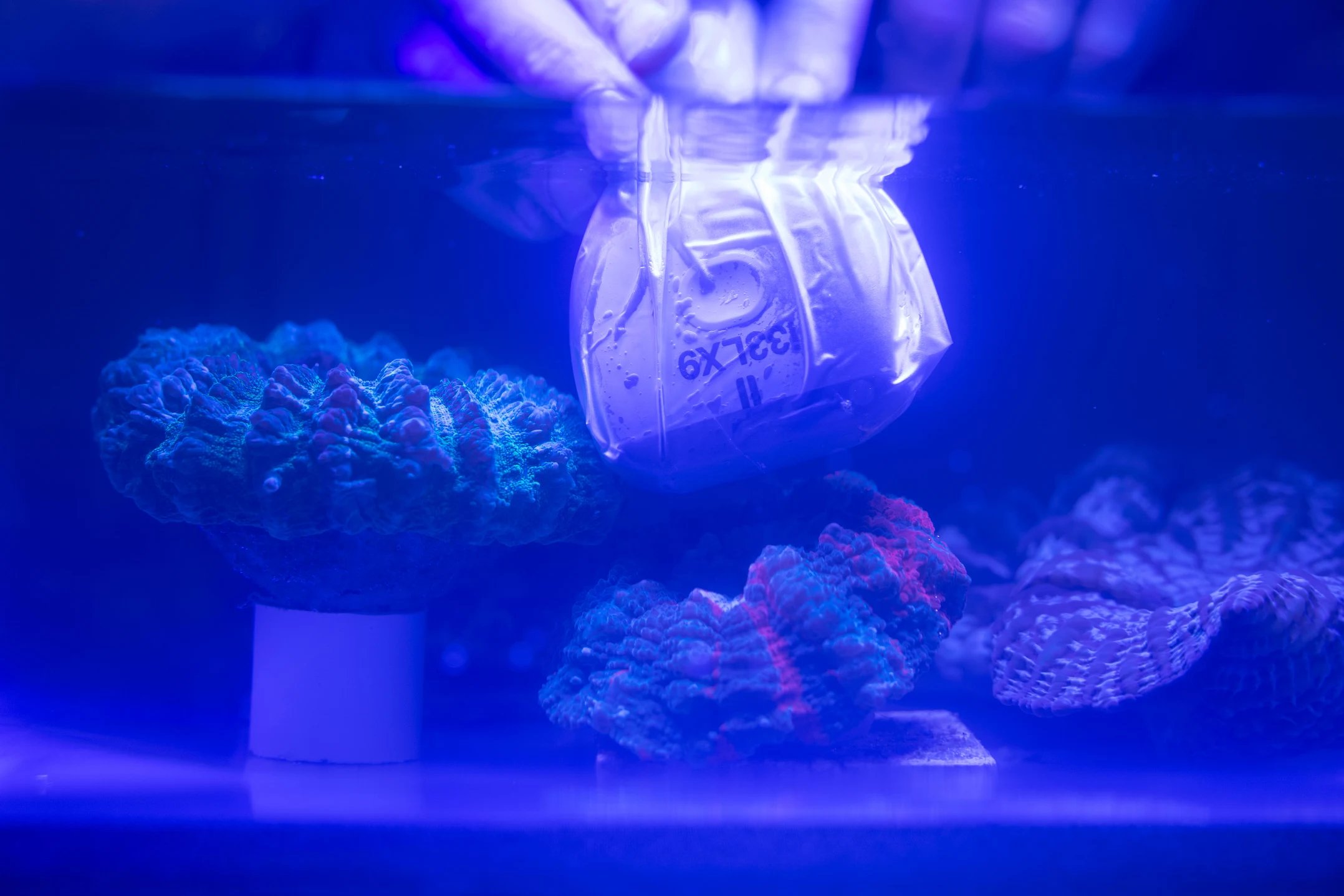
The announcement of the successful trial has drawn widespread attention from environmental technologists and corporate leadership alike, highlighting the growing intersection between commercial research and ecological preservation.
"This experiment demonstrates that imaging innovation has the power to show us what was previously unseen, creating a deeper understanding of the world around us," said Peter Bragg, Canon EMEA Sustainability and Government Affairs Director, in an official corporate statement. "By unlocking this knowledge, we hold a fundamental belief that imaging has the power to transform the world."
Marine biologists and conservation partners have similarly emphasized the practical utility of the tool in field operations. By replacing destructive dissection with rapid, painless acoustic scans, field researchers can evaluate entire colonies in situ. This capability reduces the anthropogenic stress placed on already vulnerable populations during scientific monitoring phases, allowing conservation groups to prioritize the health and longevity of broodstock corals.
Broader Environmental Context: The Global Coral Crisis
The deployment of advanced diagnostic tools occurs against a backdrop of escalating environmental distress for global marine ecosystems. In late autumn, the International Union for Conservation of Nature (IUCN) published a sobering global assessment warning that approximately 96 percent of the Earth’s coral reef ecosystems face an extremely high risk of collapse within the next fifty years.
The primary driver behind this systemic decline is anthropogenic climate change, which has triggered unprecedented oceanic heatwaves and prolonged marine warming events. Elevated water temperatures induce coral bleaching, a phenomenon where stressed corals expel the symbiotic algae living within their tissues, leading to starvation, widespread mortality, and structural degradation of the reef framework.
The loss of coral reefs carries profound ecological and economic consequences. Often described colloquially as the "rainforests of the sea," coral reefs occupy less than one percent of the ocean floor yet provide essential habitat for an estimated 25 percent of all marine species. When coral reefs suffer mass mortality, the complex reef architecture degrades into algae-dominated rubble zones incapable of supporting the same level of biodiversity. This ecological shift adversely impacts global fisheries, coastal protection against storm surges, and the livelihoods of millions of people worldwide who depend on reef ecosystems for sustenance and economic security.
Implications for Future Conservation and Restoration Strategies
Understanding the precise reproductive timing of corals is a fundamental requirement for effective active restoration. Many stony corals rely on synchronized mass spawning events, releasing millions of eggs and sperm into the water column simultaneously based on environmental cues such as lunar cycles and water temperature.
Historically, predicting these narrow reproductive windows has relied on historical averages and generalized field observations, often resulting in missed opportunities to collect viable gametes for ex-situ breeding programs, larval propagation, and cryopreservation.
The incorporation of medical-grade ultrasound scanning into marine conservation pipelines introduces a new level of precision to these efforts. By accurately detecting gametogenesis—the development of eggs—months or weeks in advance, conservationists can optimize their fieldwork schedules. Researchers can monitor captive and wild broodstock continuously, ensuring that restoration facilities are fully prepared to collect, fertilize, and nurture coral larvae during optimal reproductive windows.
While ultrasound technology alone cannot reverse the trajectory of ocean warming and acidification, it provides scientists and restoration practitioners with an essential diagnostic instrument. By bridging the gap between advanced medical imaging and marine ecology, the initiative exemplifies how cross-disciplinary innovation can be harnessed to protect endangered natural resources and improve the efficacy of global biodiversity preservation efforts.