
The Anatomy of Discovery: How a Hidden Heart Structure Is Redefining Stroke Prevention

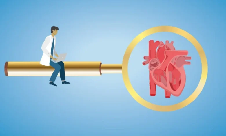
For more than a century, the medical community has operated under the collective assumption that the human heart is a fully mapped territory. Since the foundational anatomical works of Andreas Vesalius and the meticulous documentation provided by Henry Gray, physicians have been trained to view the heart’s chambers, valves, and vessels as fixed, well-understood entities. Medical students memorize these structures, and surgeons operate within their perceived boundaries, often treating the heart as a static architectural masterpiece rather than a biological landscape still capable of yielding new findings. This perspective has led to a modern scientific climate that prioritizes the frontiers of genomics, artificial intelligence, and molecular precision medicine, often at the expense of gross anatomy. However, recent findings suggest that our understanding of the cardiovascular system remains incomplete, with significant clinical implications hiding in plain sight.
The Rediscovery of Gross Anatomy
The paradigm shift began not in a high-tech laboratory, but within the silent, preserved specimens at the Jesse E. Edwards Registry of Cardiac Disease in St. Paul, Minnesota. In 2004, Dr. Subramaniam C. Krishnan, a cardiac electrophysiologist, began an independent study of these hearts. The registry, founded by the esteemed cardiac pathologist Dr. Jesse E. Edwards, was built on the premise that today’s "normal" specimen serves as the foundation for tomorrow’s diagnostic inquiry.
While examining these preserved specimens, Dr. Krishnan observed a distinct anatomical configuration within the atrial septum—a small, pouch-like structure opening into the left atrium, typically measuring between 8 and 11 millimeters in depth. Despite the heart being one of the most rigorously studied organs in medical history, this feature had evaded formal scientific classification. By 2010, following a systematic investigation of the registry’s collection, Dr. Krishnan and his colleagues published the first formal description of the left atrial septal pouch (LASP).
Prevalence and Physiological Significance
Subsequent clinical research has confirmed that the LASP is not a rare anomaly. Large-scale imaging studies suggest that this structure is present in approximately one-third of the adult population. The fact that a structural feature existing in 33% of the population remained unrecognized until the 21st century highlights a critical blind spot in clinical diagnostics: the tendency to stop looking once the standard anatomical baseline has been established.
The physiological significance of the LASP lies in its potential for blood stagnation. Much like the left atrial appendage—a well-known site where blood can pool and form clots in patients with atrial fibrillation—the LASP acts as a blind-ended cul-de-sac. Crucially, research has indicated that clot formation can occur within the LASP even in patients with a normal, healthy heart rhythm. This led investigators to hypothesize that the pouch could be a previously unidentified culprit in "cryptogenic" strokes, or strokes for which no clear cause can be identified.
A Chronology of Clinical Validation
The journey from an anatomical observation to a clinical trial follows a rigorous, albeit slow, trajectory common in cardiology.
- 2004–2009: Dr. Krishnan and colleagues conduct systematic studies at the Jesse E. Edwards Registry.
- 2010: The first formal description of the Left Atrial Septal Pouch (LASP) is published in the Journal of the American College of Cardiology: Cardiovascular Interventions.
- 2015–2023: Multiple observational studies identify an association between the LASP and ischemic strokes, raising questions about whether the pouch requires clinical intervention.
- September 2024: Investigators at Fuwai Hospital in Beijing report the findings of a randomized trial evaluating the transcatheter elimination of the pouch.
The Fuwai Hospital study represents a watershed moment. By utilizing septal occluder devices—originally designed for the closure of a patent foramen ovale (PFO)—researchers were able to mechanically seal the pouch. The results demonstrated a marked reduction in recurrent, MRI-detected ischemic brain lesions in patients who underwent the procedure compared to those treated with standard medical therapy alone. While medical experts emphasize that this study is not the definitive final word, it provides the first empirical evidence that direct intervention of the LASP can lead to improved patient outcomes.
Parallels with the Patent Foramen Ovale (PFO)
The trajectory of the LASP mirrors the history of the PFO, a common hole in the heart that connects the right and left atria. For decades, the PFO existed in a state of clinical limbo—recognized by anatomists but debated by clinicians regarding its role in stroke pathology. It took years of epidemiological data, refined imaging techniques, and extensive randomized clinical trials to move PFO closure into the mainstream as an accepted therapy.
The LASP is following a similar path, but at an accelerated pace, thanks in part to the advancements in diagnostic imaging that were not available when the PFO was first being debated. The integration of clinical observation with modern interventional techniques has allowed the medical community to transition from asking "What is this?" to "How do we treat it?" with unprecedented speed.
Broader Implications for Medical Science
The discovery of the LASP serves as a cautionary tale for the modern medical establishment. As healthcare systems pour billions of dollars into AI-driven diagnostic tools and genomic sequencing, there is an inherent risk of devaluing traditional clinical observation. The LASP discovery required no billion-dollar technology; it required an inquisitive mind, access to physical specimens, and the willingness to challenge the status quo.
Furthermore, the study of the LASP has already begun to influence other areas of cardiovascular care. Dr. Krishnan’s subsequent work, which explores an implant-free strategy for closing PFOs, is a direct result of insights gained while studying the pouch. By viewing the LASP as an incompletely fused PFO tunnel, researchers have hypothesized that it may be possible to induce focal biological adhesions that naturally seal these defects without the need for permanent, synthetic implants.
The Future of Anatomical Research
The medical community faces a dual challenge: maintaining the momentum of technological innovation while preserving the fundamental skills of gross anatomy and careful physical observation. The LASP is a reminder that the human body is a living science, and our understanding of it is perpetually evolving.
"Modern medicine should continue investing heavily in genomics, molecular biology, and artificial intelligence," says Dr. Krishnan. "But those advances shouldn’t persuade us that anatomy has finished teaching us. As we spend billions teaching computers to recognize patterns in the human body, we must not lose the ability to recognize them ourselves."
Ultimately, the lesson of the left atrial septal pouch is that the most transformative medical discoveries are not always found in the microscopic sequences of a genome or the high-speed processing of an algorithm. Sometimes, they are found in the overlooked corners of the human heart, waiting for an observant eye to recognize them. As medicine looks toward the future, it must remain grounded in the reality that the most significant breakthroughs may still be hiding in plain sight, within the anatomy we presumed to have mastered generations ago.
This discovery recalibrates the focus of cardiac research, encouraging a return to foundational investigations that bridge the gap between basic anatomy and clinical intervention. As researchers at institutions like Fuwai Hospital and others continue to refine the surgical approach to the LASP, the clinical landscape for stroke prevention may be permanently altered, demonstrating once again that in the field of medicine, the simplest observations often lead to the most profound impacts on patient health.