
Parasitic Invasion of the Brain: A Rare Case of Neuroschistosomiasis in a Filipino Rice Farmer

Out of nowhere, a woman’s right arm and leg started shaking uncontrollably. In the moments leading up to the experience, she appeared to be in perfect health; she reported no preceding headaches, mental confusion, or physical weakness. Yet, for approximately one minute, the right side of her body underwent intense, involuntary spasms. This sudden onset of focal seizures prompted the woman, a rice farmer residing in the Philippines, to seek urgent medical attention at a local emergency department.
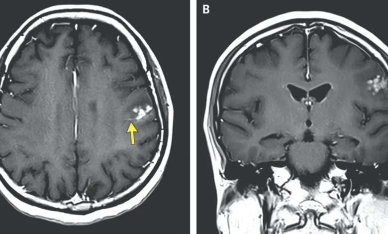
Upon her arrival, attending physicians conducted a thorough neurological examination. To their surprise, the physical exam yielded results that were entirely unremarkable; she showed no signs of localized muscle atrophy, cognitive impairment, or cranial nerve deficits. However, given the specificity of her symptoms—the localized shaking—doctors prioritized advanced diagnostic imaging to investigate potential intracranial pathology. Magnetic resonance imaging (MRI) of the brain revealed a startling finding: a cluster of bright, distinct nodules surrounded by localized fluid accumulation located in the left frontal lobe. The clinical team immediately suspected a parasitic etiology, specifically targeting the possibility of neuroschistosomiasis.
Clinical Diagnosis and Pathogenesis
The case, detailed in a report published in the New England Journal of Medicine, highlights the rare but dangerous neurological complications associated with Schistosoma, a genus of trematode flatworms, or "blood flukes." While schistosomiasis typically affects the liver, intestines, or urinary tract, neuroschistosomiasis occurs when the parasitic eggs are deposited in the central nervous system, triggering an inflammatory response that leads to seizures, weakness, or increased intracranial pressure.
In the Philippines, the primary causative agent for this infection is Schistosoma japonicum. Despite the geographical misnomer in its name—it was officially eliminated in Japan decades ago—the parasite remains endemic in parts of the Philippines, China, and Indonesia. The infection pathway is remarkably complex and highly efficient at bypassing the human body’s standard immunological defenses.
The Diabolical Life Cycle of S. japonicum
The lifecycle of the blood fluke is a masterclass in evolutionary adaptation. The cycle begins when eggs are shed into the environment via the feces or urine of an infected mammalian host, such as humans, cattle, or pigs. When these eggs reach freshwater, they hatch into miracidia—a free-swimming larval stage. These larvae must locate a specific intermediate host, the amphibious snail, to survive.
Once inside the snail, the parasite undergoes a period of asexual reproduction, transforming through two distinct developmental stages. Eventually, the parasite emerges from the snail as a cercaria—a free-swimming larva equipped with a forked tail designed for penetration. These cercariae are highly motile and seek out mammalian skin. Upon contact, they use specialized enzymes to tunnel through intact skin, entering the vascular system.
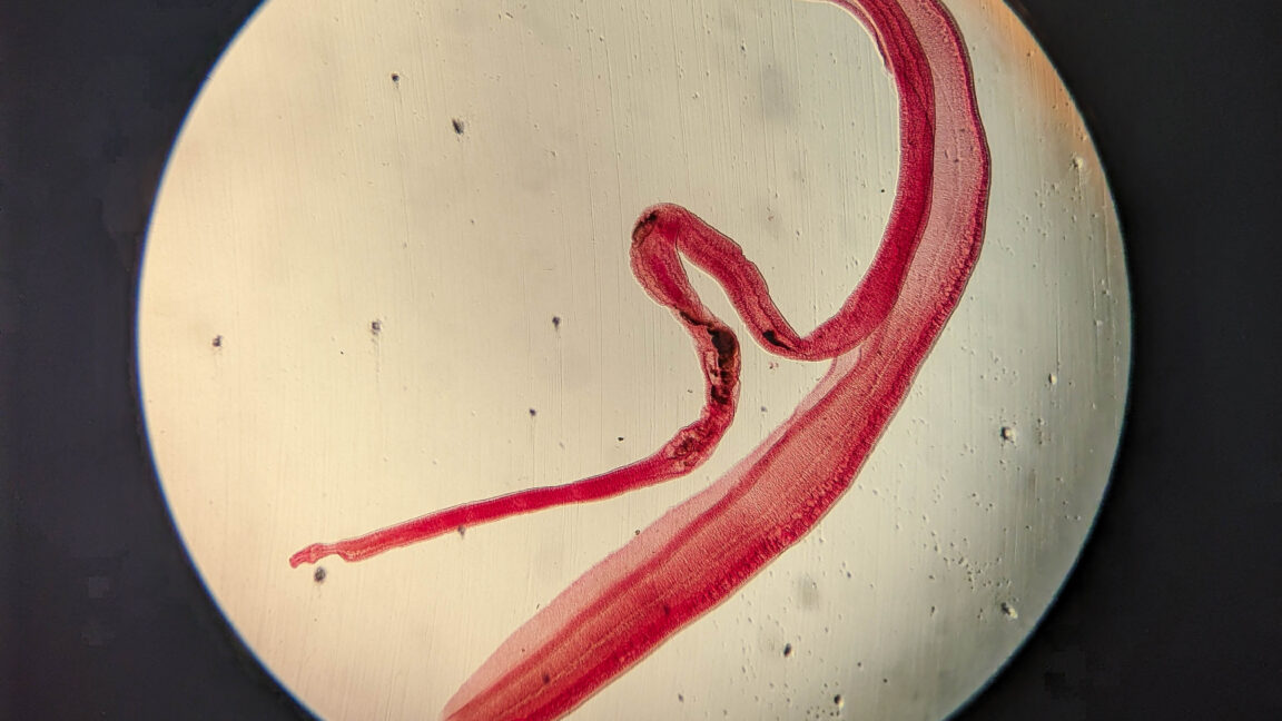
Once inside the host, the larvae migrate through the heart and lungs before settling in the intrahepatic portal blood vessels. Here, they mature into adult worms. In a unique biological pairing, the slender female resides within a specialized groove on the body of the thicker male, effectively "cuddling" for the remainder of their adult lives. These pairs reside in the mesenteric veins, where the female releases thousands of eggs daily. While many eggs exit the body, a subset can be carried by the bloodstream to the brain or spinal cord, resulting in the localized nodular lesions observed in the Filipino farmer’s MRI.
Timeline of the Clinical Incident
The patient’s experience follows a classic, albeit alarming, progression for neuro-parasitic infection. While the exact moment of exposure is difficult to pinpoint due to the long incubation period of schistosomiasis, the clinical timeline observed by her physicians was relatively acute:
- Pre-Symptomatic Phase: The patient remained asymptomatic for a period likely spanning weeks or months, during which the larvae were migrating through her vascular system.
- The Incident: The focal seizure lasted for approximately 60 seconds. This is consistent with activity in the motor cortex of the left frontal lobe, which controls the movement of the right side of the body.
- Emergency Presentation: Upon arrival at the hospital, the patient’s status was stable, indicating that the seizure was an isolated event rather than an ongoing systemic collapse.
- Imaging and Confirmation: The MRI provided definitive visual evidence of granulomas—the body’s immune system walling off the parasitic eggs. This inflammatory reaction is what causes the surrounding fluid and the subsequent electrical misfiring in the brain that leads to a seizure.
Supporting Data and Public Health Context
According to the World Health Organization (WHO), schistosomiasis remains a significant public health challenge in tropical and subtropical regions. It is classified as a Neglected Tropical Disease (NTD), affecting millions of people who live in rural communities lacking adequate sanitation and safe water. In the Philippines, the prevalence of S. japonicum has historically been a barrier to rural development, particularly in agricultural sectors where farmers are in frequent contact with potentially contaminated irrigation water.
Research data indicates that while neuroschistosomiasis is reported in less than 5% of all schistosomiasis cases, it carries a high morbidity rate if left untreated. The diagnosis is often challenging because the symptoms can mimic tumors, tuberculomas, or other neurological conditions. The case published in the NEJM serves as a critical reminder to clinicians in endemic areas to consider parasitic infections when patients present with sudden-onset focal seizures, even in the absence of other systemic symptoms like fever or liver enlargement.
Clinical Implications and Management
The management of this case involves a dual-pronged approach: the administration of anti-parasitic medication, typically praziquantel, and the use of corticosteroids to manage the inflammation around the nodules. Praziquantel works by increasing the permeability of the worm’s cell membrane, causing paralysis and death of the parasite, while steroids are essential to prevent further neurological damage caused by the inflammatory response of the brain tissue as the parasites die.
Medical experts involved in the study emphasize that the patient’s prognosis is generally favorable provided the infection is identified early. The nodules, while alarming on an MRI, are treatable, and the seizures can usually be managed with standard anti-epileptic drugs until the inflammation subsides. However, the case underscores the necessity of improved public health infrastructure, specifically the control of snail populations and the implementation of better sanitation facilities in rice-farming communities.
Broader Impact: The "One Health" Challenge
The case of the Filipino rice farmer illustrates the intersection of human, animal, and environmental health—a concept known as "One Health." Because S. japonicum is a zoonotic parasite (meaning it infects both humans and animals), controlling the disease is not merely a matter of treating human patients. It requires an integrated approach that includes:
- Water Management: Improving the safety of water used for agriculture and domestic purposes to prevent cercarial penetration.
- Snail Control: Targeted efforts to reduce the habitat of the intermediate snail host.
- Livestock Treatment: Regular deworming of farm animals, which often serve as reservoirs for the parasite.
- Public Education: Informing agricultural workers about the risks of skin exposure to stagnant water and the importance of wearing protective gear.
This case, while rare, serves as a poignant reminder that globalization and climate change continue to influence the prevalence of parasitic diseases. As farming practices evolve and rural populations expand into previously undisturbed ecosystems, the risk of zoonotic transmission remains a persistent concern. For the patient in this report, the prompt application of advanced imaging and the medical community’s familiarity with local endemics ensured a path to recovery. Her story stands as a testament to the importance of global medical collaboration and the ongoing effort to manage the silent, microscopic threats that coexist within our ecosystems.
Related posts: